


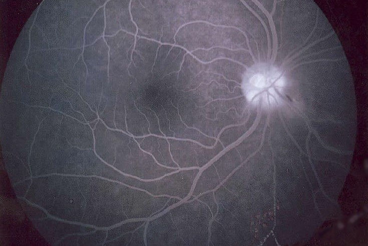
A study comparing five different optical coherence tomography angiography (OCTA) systems with fluorescein angiography (FA) found FA still obtains the best results to visualise microaneurysms, which is crucial for the diagnosis of diabetic retinopathy (DR).
Detecting the presence of microaneurysms (MAs) is an important, early pathologic sign to assist the diagnosis and staging of DR, said study researchers from the University of Milan. Historically, widely clinically available colour fundus photography hasn’t been able to distinguish MAs from haemorrhages, with MAs appearing as little red dots. For this reason, fluorescein angiography (FA) has been considered the gold standard for MAs detection, visualised as hyperfluorescent dots on early phases. OCTA, however, offers the opportunity to visualise different retinal capillary plexus and the choroidal vasculature without the need for dye injection, said researchers.
The Milan-based study enrolled 15 patients with DR (20 eyes). The patients were imaged with FA and five OCTA devices - prototype Spectralis OCTA (Heidelberg Engineering), prototype PlexElite (Zeiss), RTVue XR Avanti (Optovue), AngioPlex (Zeiss) and DRI OCT Triton (Topcon) - on the same day. For all OCTA devices, a 3×3 volume scan pattern was performed. MAs were evaluated in the superficial capillary plexus (SCP) and in the deep capillary plexus (DCP).
Analysing the results, researchers found FA resulted in finding a significantly higher number of MAs compared with OCTA devices. Spectralis OCTA obtained a significantly higher number of MAs compared with PlexElite, RTVue XR Avanti, AngioPlex and DRI OCT Triton (p<0.0001). PlexElite and AngioPlex showed a greater number of MAs in the SCP, Spectralis OCTA, RTVue XR Avanti and DRI OCT Triton in the DCP. Higher sensitivity (43.3%) but lowest specificity (54.4%) was observed for Spectralis OCTA compared with other devices. The higher specificity (78.5%) and positive predictive value (83.3%) were observed for DRI OCT Triton.
“Comparing five different OCTA devices and traditional FA in the ability to detect retinal MAs, FA appeared to be the best imaging modality to identify retinal MAs,” said co-author Dr Federico Corvi from the department of biomedical and clinical science at Luigi Sacco Hospital. “The Spectralis OCTA was able to detect more MAs compared to other devices, likely due to the higher number of B-scans in the scanned area as well as due to the higher number of repeated B-scans.” Though different OCTA devices showed a higher number of MAs in the SCP while others showed more in the DCP, he said. “The high variability between OCTA devices should be taken into account for future clinical trials as in clinical practice.”
The study was published by the British Journal of Ophthalmology.