


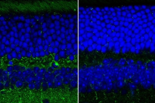
US researchers have developed an imaging technique measuring both the thickness and the textures of the retinal layers, potentially offering an early warning system for Alzheimer’s disease.
“Previous research has seen a thinning of the retina in Alzheimer’s patients, but by adding a light-scattering technique to the measurement, we’ve found that the retinal nerve fibre layer is also rougher and more disordered,” said study lead Dr Adam Wax, professor of biomedical engineering at Duke University in North Carolina.
Using angle-resolved low-coherence interferometry (a/LCI), which measures the angles of scattered light to gather information about the tissue’s structure, the researchers found that the topmost layer of neurons in the retina of a mouse model of Alzheimer’s disease exhibit a change in their structural texture. Combining this information with data on thickness layer changes, derived from Optical Coherence Tomography (OCT) imaging, this provides a new measurement that could prove to be a more easily accessible biomarker of Alzheimer’s, said Prof Wax.
“The a/LCI measurements complement the thickness measurements to improve the potential utility of more quantitative biomarkers for Alzheimer's,” said graduate student Ge Song. “You can’t get textural and structural information about the retina with OCT alone. You need both imaging modalities. That’s the key innovation.”
Study results were published in Scientific Reports. Researchers are now working to incorporate this added ability into a low-cost OCT system being developed by Prof Wax through university spinoff company Lumedica.