


One of the great things about our speciality is that we have so much wonderful technology at our disposal. One of the terrible things about our speciality is that we have so much wonderful technology at our disposal. Wait, what?
The thing is, for every potentially helpful new piece of technology we have to do the business case, and that applies whether in public or private practice. You have to decide how much value the new piece of equipment will add, how often you’ll use it and what the financial implications are. Only then can you consider making a purchase.
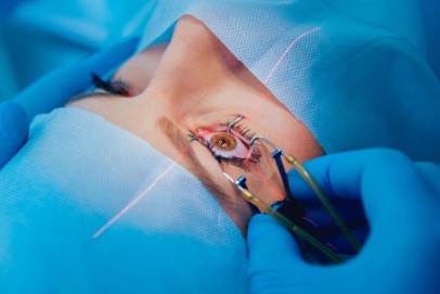
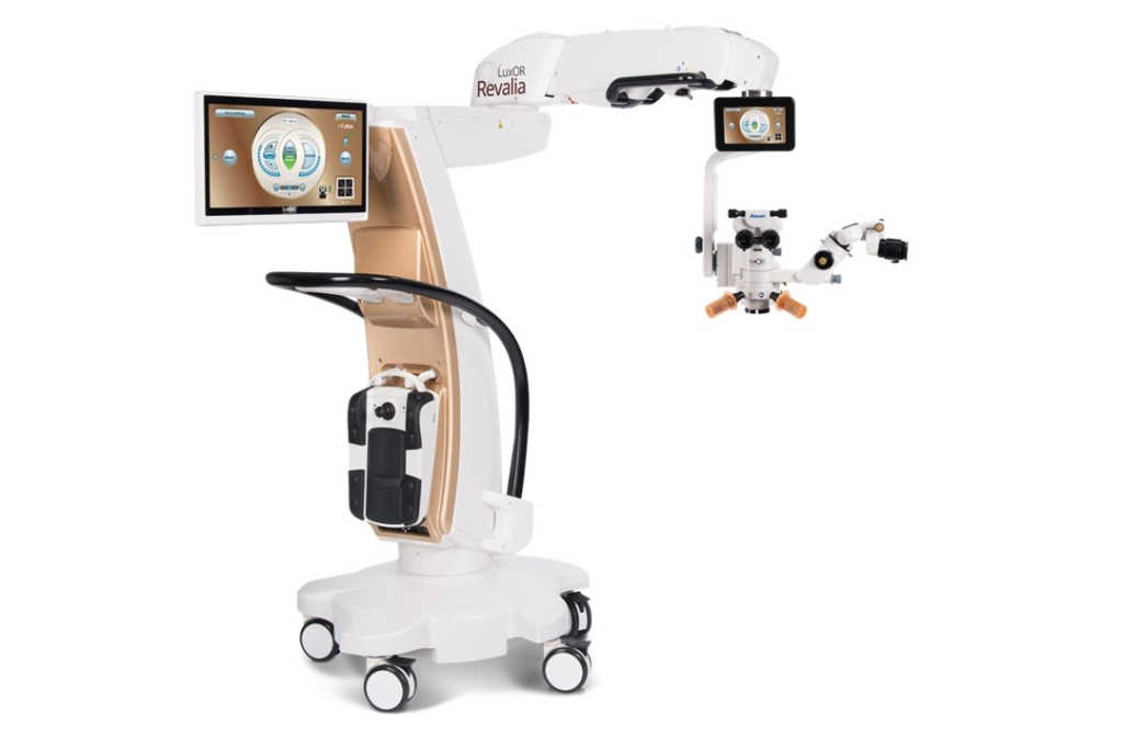
Late last year I upgraded my operating microscope. Modern microscopes, like modern cars, all work well and there is often not a lot to distinguish one from another. I had been using a well-known brand of microscope and it worked perfectly well. My reason for investigating new options was to improve my toric outcomes. Like many surgeons, I have used toric implants sparingly: they cost a lot more than spherical lenses and aren’t funded by insurance companies. Anyone paying good money for a premium lens quite reasonably expects a premium outcome.
Most of us mark the limbus prior to surgery with the patient upright at a slit lamp. But this can be erratic – the eyelid got in the way, the patient blinked repeatedly, the surgical marker pen was too thick. So it can be a frustrating start to the procedure and not that accurate. Enter the era of the digital overlay. At the time of biometry, the Verion Image Guidance System registers iris and limbal details and the data is transferred to my new Luxor Revalia ophthalmic microscope. At the start of surgery, the patient’s name, correct eye and toric axis are all visible through the microscope’s right-hand eyepiece. The surgeon and scrub nurse confirm that registration is complete and the procedure can begin. This all takes a matter of seconds. Somewhat crucially the nurses say that data capture and transfer is a simple process – technology has to be user friendly for all members of the team!
The Verion system also integrates perfectly with the Centurion phacoemulsifier. The overlay changes as the surgeon advances through the phaco settings. I use the incision marker, capsulorrhexis guide and toric axis marker. These settings can be customised to suit each surgeon’s preferences. I have found it quite interesting to see how variable the rotation of each eye can be as patients move from upright to supine. It seems I haven’t always been placing my incisions exactly where I had thought!
Another advantage of the new microscope is the light source. Quite anecdotally, patients with my previous microscope would almost always comment on the brightness of the light when first rotated into position, despite it being on its lowest setting. The new microscope is much better tolerated, and it is very much the exception for a patient to remark on the harshness of the light. With this system, the light settings are also easily customised, eg. more coaxial light for cataract surgery and more oblique light for pterygia. I like to have a few favourite settings saved as I’m not one to fiddle endlessly with equipment during a procedure. The depth of field and red reflex are also excellent.
Future audits will show how much difference these digital overlays are making, but in the meantime, it feels good to know I’m operating with a greater degree of precision.
Dr Brian Kent-Smith is a consultant ophthalmologist at Eye Specialists in Northland, New Zealand, and former chair of the RANZCO NZ Branch. He has no affiliations with Alcon or any other ophthalmic products company.