


Heidelberg Engineering is reintroducing its HRT3 Rostock Cornea Module (RCM) offering in-vivo corneal confocal microscopy, to support clinicians managing corneal diseases.
The unmet clinical need for high-resolution in-vivo corneal microscopy paved the way for the decision, said Erich Bangert, Heidelberg Engineering’s vice president. “With its ability to investigate the cornea at the cellular level, HRT3 RCM will empower clinicians in fast-growing clinical areas such as dry eye disease, infectious corneal diseases, corneal dystrophies, diabetic neuropathy and refractive and corneal treatment surgery.”
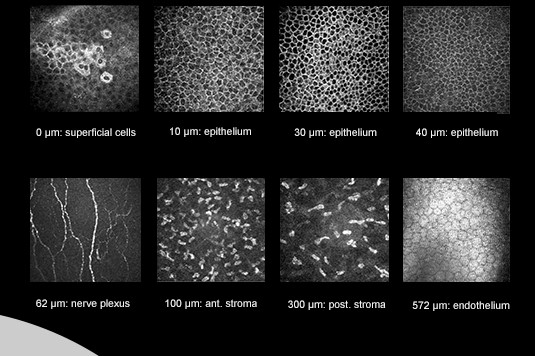
The microscope scans the cornea with a field of view of up to 400x400μm, resulting in high-resolution images allowing the user to navigate through all corneal layers, including the subdifferentiation of various epithelial cell layers; identify keratocytes subpopulations; and visualise the corneal sub-basal nerve plexus, said Bangert.
The HRT3 RCM comes with a headrest for corneal assessment and Heidelberg’s Heyex 2 image management software for streamlined workflows, including automated report generation, archiving and drag-and-drop exports for paperless data sharing, said the company.
The HRT3 reintroduction is scheduled for early to mid-2020.