Human stem cells restore primate’s sight
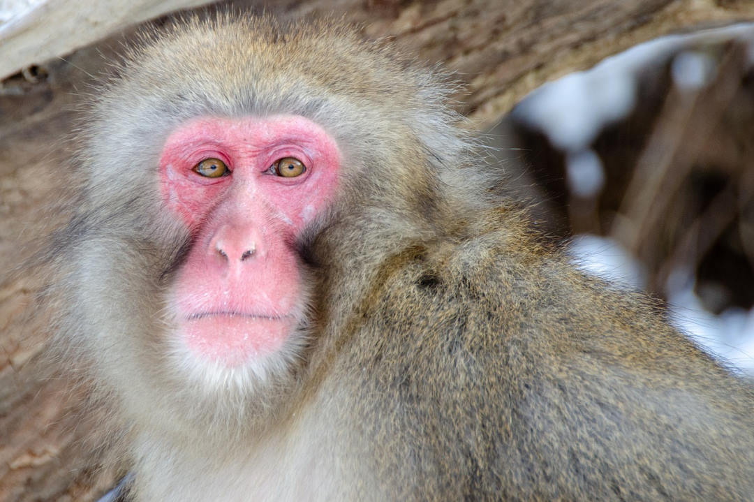
Researchers in Japan restored a Japanese macaque’s macular hole (MH) using a human stem cell graft, which improved visual function.
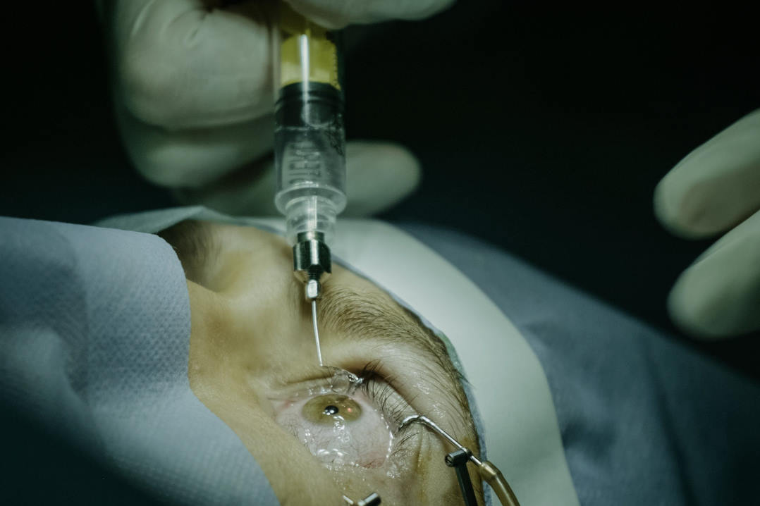
Writing in Stem Cell Reports, a team led by Dr Michiko Mandai, Laboratory for Retinal Regeneration, RIKEN Center for Biosystems Dynamics Research, Kobe, described the implantation of human embryonic stem cell (hESC)/induced pluripotent stem cell-derived retinal organoid (RO) sheets over the macaque’s MH. No silicone oil tamponade was used, since intraoperative OCT showed the graft looked fixated under the retina in the whole circumference of the MH, they noted.
The graft contributed to macular hole closure with continuously integrated tissue containing rod, S- and L/M-cone photoreceptors and improved visual function, said researchers. However, four months post-surgery, they observed mild rejection of the patch, which may have been due to the cross-species nature of the transplant, Dr Mandai told Live Science. "Transplantation of human tissue to a human would have less risk of immune response," she said.
This approach simplifies an autologous retinal transplantation procedure, with the advantages of no need for harvesting the peripheral retina or preparation of different sizes of sheets, including the one for a large MH; plus, the hESC graft includes cone photoreceptor precursor cells, they said. Further studies are needed to validate the functional advantage of the pluripotent stem cell-derived retina, including the synaptic connectivity with and protective effect for host retinal cells, they added.


