


Using a new imaging technique, University of Melbourne researchers have discovered the immune cells protecting the cornea are T cells, not dendritic (or Langerhans) cells, as was previously thought.
Led by Associate Professor Laura Downie, Dr Holly Chinnery and Professor Scott Mueller, the research team used a non-invasive imaging approach they termed ‘functional in vivo confocal microscopy’ (Fun-IVCM), to observe T cells moving around quickly and interacting with other cells and nerves in the outermost layer of the cornea. “We also captured different cell dynamics in response to contact lens wear and in allergic eye disease and quantified how these behaviours are modulated by drug treatments,” said A/Prof Downie.
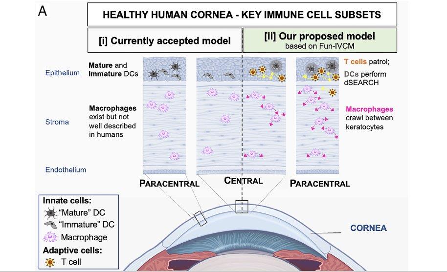
The currently accepted model of the principal immune cell subsets in the healthy human cornea, involving ‘mature’ and ‘immature’ dendritic cells in the epithelium (as imaged using traditional static in vivo confocal microscopy (IVCM) and macrophages in the stroma). [ii] A proposed role for adaptive immune cells (T cells) in homeostatic human corneal immune surveillance. Credit: University of Melbourne
Writing in Proceedings of the National Academy of Sciences, authors said Fun-IVCM allows researchers to study the interface between nerves and immune cells in an intact peripheral sensory tissue, allowing them to assess how exogenous factors and disease may disrupt the perception, integration and responsiveness of cell subtypes to different challenges. “These findings reshape our understanding of the distinct immune cell subsets in the human cornea and how they respond to different stimuli,” said A/Prof Downie.