What to expect when your patient is expecting
Anyone who has been pregnant or lived with someone who was pregnant knows that being expectant comes with its fair share of surprises – good and bad. Changes in the release of hormones such as oestrogen, progesterone, relaxin and others incite a chain reaction with subsequent changes in vascularity, fluid retention and tissue remodelling. These changes may also have effects on how the eyes look and function, too. Let’s take a look at some of the eye-related clinical considerations and possible pathologies during pregnancy.
Pigmentation
An increase in the release of oestrogen, progesterone and melanocyte-stimulating hormones during pregnancy can lead to an increase in pigmentation in the body, including periorbitally1. As clinicians, we need to be able to differentiate iatrogenic causes of hyperpigmentation (for example, use of prostaglandin analogues) from those related to pregnancy.
Vision
Hormonal modulations also lead to swelling in the lens and cornea, most often creating a myopic shift (although sometimes it may be hyperopic or astigmatic). This is why procedures addressing refractive correction (such as LASIK) are contraindicated during pregnancy.
When screening women of child-bearing age for laser eye surgery, we must enquire about pregnancy or breastfeeding, says Dr Lana Del Porto, a Melbourne-based ophthalmologist specialising in cataract, refractive and strabismus surgery. “It’s important we have a very accurate understanding of the patient’s refractive error and that the prescription is not changing when planning laser eye surgery. For this reason, we do not offer it when the patient is pregnant or breastfeeding. Instead, we invite them to return for an assessment, generally six weeks after cessation of breastfeeding.”
The ocular surface
Increasing gestational age is associated with changes to tear-film physiology and increased incidence of dry eye in pregnant women.
Pregnancy leads to several ocular surface changes, which are further exacerbated by existing pathology, says Dr Stuti Misra, an optometrist-scientist and senior lecturer at the University of Auckland’s Department of Ophthalmology. “Dry eye disease is known to occur throughout pregnancy but increases with gestational age2. Common assessments including tear breakup time and Schirmer test are reported to worsen in the third trimester of pregnancy3.”
There is conflicting evidence on the cause of this association. Hormonal modulations to oestrogen/prolactin/testosterone levels and immunoarchitectural changes to the lacrimal gland have been postulated to play a role4. Similarly, there is no consensus on whether pregnancy is related to evaporative, aqueous deficient, or mixed dry eye5,2. Following the DEWS II dry eye sub-group diagnostic tests, including a dry eye questionnaire, non-invasive tear breakup time, Schirmer test and identification of meibomian gland disorder (eg, through meibography/meibomian gland assessment) can help us adequately assess and manage the presentation of dry eye in our pregnant patients5,6.
Keratoconus
Pregnancy can also be considered a risk factor for keratoconus progression, affecting not only corneal topography, thickness measurements and curvature, but also biomechanical properties such as corneal hysteresis and corneal resistance factor7. Again, the mechanisms behind these changes are unclear; they could be related to lower thyroxine levels, changing oestrogen levels, or increasing relaxin levels (changing synthesis of MMPs and TIMPs)8. Interestingly, there is some evidence there may be some partial recovery after cessation of breastfeeding9, indicating that clinicians should wait for stabilisation of corneal parameters before determining a management approach. “I had a patient last year who had recently moved to Australia from Canada,” says Dr Del Porto. “She had keratoconus. Her corneal tomography showed significant worsening of her keratoconus compared to three years prior. During that time, she had been pregnant twice.”
Keratectasia
Dr Misra points out that a scoping review noted the tendency of pregnant patients to develop iatrogenic keratectasia several years after receiving Lasik, with or without preoperative risk factors. “Importantly, the ectasia may re-occur in second or third pregnancies. However, most of the related ocular symptoms are likely to persist postpartum10. A few studies have also shown steepening of topography, a decline in spherical equivalent refraction and an increase in astigmatism11.” So, in addition to standard refraction for pregnant patients, it may be of benefit for optometrists to conduct a complete dry eye disease work-up, along with corneal topography, Dr Misra suggests.
Intraocular pressure and glaucoma
Studies have shown intraocular pressure (IOP) can decrease by up to 20% during pregnancy12. There are not enough randomised controlled trials to understand the physiology behind this change, but it has been hypothesised it could be multifactorial – attributable to increased aqueous outflow, decreased episcleral venous pressure (from an overall decrease in systemic vascular resistance) and decreased scleral rigidity due to increase in tissue elasticity (from oestrogen and progesterone release)13. This is a genuine decrease in IOP and not artefactual, so normal IOP cut-offs for our pregnant patients with glaucoma and ocular hypertension should be applied.
Conversely, there is some evidence that a history of pregnancy that results in delivery (especially three or more) can be associated with an increased incidence of open angle glaucoma14. This has been suggested to be related to high oestrogen levels during pregnancy; transient events during labour (including systemic hypotension and decreased ocular perfusion because of bleeding) inducing glaucoma-like changes in the optic nerve; oxytocin levels during labour inducing capillary constriction and decreasing aqueous outflow; stress during labour causing the release of adrenaline and noradrenaline that lead to an increase in IOP; and the valsalva reflex during labour producing intermittent increases in IOP.

Uveitis
It is hard to generalise about pregnant women who have uveitis associated with autoimmune conditions (Behcet disease, idiopathic, sarcoidosis, Vogt-Koyanagi-Harada-associated, juvenile idiopathic arthritis etc.) as there are issues with sample size and limited studies when it comes to the literature. From what is available, though, uveitis appears to worsen in the first trimester, be less active later on, and increases postpartum, with an increased risk of uveitis flare within six months of delivery15,16. More than 50% of patients in a case series experienced a uveitis episode in the first six months postpartum16.
Neuro-ophthalmic presentations
Multiple sclerosis is another autoimmune inflammatory condition that presents a similar situation: most cases improve during pregnancy, but attacks (including episodes of optic neuritis) can increase in the first few months postpartum17.
Idiopathic intracranial hypertension can be exacerbated during pregnancy, especially as maternal weight gain increases with increasing gestational age18. Pituitary adenomas are another pathology that can progress during pregnancy, with the classic bitemporal visual-field defect becoming more prominent or deepening/expanding19. For each of these presentations we should follow our pregnant patients a little more frequently, with appropriate visual field and optical coherence tomography performed as indicated.
During the postpartum period, pituitary apoplexy/Sheehan syndrome is a neuro-ophthalmic presentation to watch out for. With the significant blood loss associated with labour and the postpartum period, a large haemorrhage can decrease perfusion of the pituitary gland. This infarct leads to swelling of the pituitary gland, which leads to compression of the visual pathway and symptoms such as sudden headache, diplopia, or vision loss20.
Posterior eye and visual pathway
Pregnancy can be considered an immune condition (since there are genetic differences between the mother and foetus). This can alter immune functionality and in some instances be related to toxoplasmosis reactivation21.
Preeclampsia is a condition that may develop in women who are more than 20 weeks pregnant. Blood pressure rises above 140/90 (this does not need to be sustained) and there is proteinuria. Preeclampsia exists on a spectrum, with a variant being HELLP (haemolysis, elevated liver enzymes and low platelets) syndrome, and more end-stage presentations including eclampsia and posterior reversible encephalopathy syndrome (PRES). Each of these presentations can cause visual changes such as blurred vision, photopsias, scotomas, diplopia and, in the case of PRES, even cortical infarcts that lead to visual field defects and cortical blindness22. The treatment for these presentations targets the eclampsia; if your patient’s vision is affected and they have a positive history for one of these presentations, then you must take a multidisciplinary approach and contact their obstetrics team.
This spectrum of presentations can also lead to ocular signs such as bilateral serous retinal detachments, or retrograde retinal nerve fibre layer/ganglion cell damage. Since first observation could even be in the postpartum period, it is important to get a thorough history of the patient’s pregnancy, where relevant. If there is a positive history, once again, a multidisciplinary approach with involvement from general practice (including vascular workup), obstetrics and endocrinology could be indicated.
The exact pathophysiology of the serous detachments is unknown, but current knowledge points to a link to the higher choriocapillaris vascular density in pregnant women. When there is a comorbidity of hypertension, there can be formation of microthrombi in the choroidal vasculature, and this ischaemic environment causes a breakdown of the blood-retina barrier and reduced resorption of subretinal fluid, ultimately leading to a serous retinal detachment23.
Vascular changes
During pregnancy cardiac output and systemic volume increases by 30–50%, blood vessels swell and become more friable and there is a decrease in fibrinolytic activity, creating a hypercoagulable state. This can lead to retinal arterial and venous occlusion during pregnancy. This is especially relevant for patients with pre-existing thrombophilic conditions or other underlying disease (such as preeclampsia/thrombotic thrombocytopenic purpura). The resulting increase in venostasis means relevant patients are often prescribed prophylactic anticoagulants24.
Diabetes
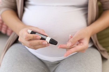
Worldwide, there is an increased incidence of diabetes, obesity and increased maternal age at first pregnancy. This has created an increasing likelihood of comorbidity of pregnancy and diabetes. It is important to note that a patient with gestational diabetes is less likely to develop diabetic retinopathy (DR); rather, it is the patients with existing DR who we should watch more closely as their retinopathy is more likely to progress. This is because the haemodilution associated with pregnancy leads to a more hypoxic/ischaemic retinal environment, which can exacerbate the DR. The risk factors for progressing DR during pregnancy are poor glycaemic control, comorbid hypertension, and degree of retinopathy pre-pregnancy25. Interestingly, during pregnancy HbA1c is no longer the gold standard method to gauge glycaemic control, since the increased renal clearance during pregnancy makes HbA1c artificially low. Instead, clinicians may ask to review blood glucose log sheets, and obstetricians will make decisions based on the glucose tolerance test26.

Patients with non-proliferative DR (NPDR) can experience up to 50% progression, with some regressions being observed postpartum. Severe NPDR transitions to proliferative diabetes in 5-20% of cases and there is progression of up to 45% in those with existing proliferative DR (PDR)27. So, depending on the severity of your pregnant patient’s pre-pregnancy level of DR, comorbidities and glycaemic control, they may need to be reviewed every trimester, or even monthly.
Guidelines for treating DR
Guidelines based on the available evidence on DR and pregnancy recommend treatment in pregnant patients only when their DR has progressed to PDR. This is because the treatment paradigms for pregnant women with DR are different. Since anti-VEGF therapy can alter placental growth factor it must be avoided in pregnancy, especially during the first trimester. Beyond that, it will be a risk-benefit conversation between the ophthalmologist and the patient24. Additionally, bevacizumab (Avastin) has a prolonged systemic effects profile. There are limited data on this matter, but it is interesting to note that in a study with three patients who received intravitreal bevacizumab therapy during pregnancy, all of the resulting toddlers reached developmental milestones appropriately during infancy28. Another notable finding in this review is that more than 50% of the women receiving intravitreal anti-VEGF were only discovered to be pregnant after receiving the injection28. This raises the question of whether pregnancy tests should be a routine consideration in relevant patients before administering intravitreal anti-VEGF.
Intravitreal steroid injection also risks systemic absorption, so typically the treatment of choice during pregnancy is retinal laser therapy27.
One way this knowledge can improve our practice is by asking relevant patients with DR if they are planning to be pregnant in the next year. If they are, it is better to pursue treatment of the DR and get it under control before they are pregnant, since we know it is likely to progress. For example, it would be clinically justified to treat a patient with severe NPDR who is planning to become pregnant in the next 12 months. It could also be a motivation to better define the full extent of DR (since some peripheral neovascularisation is undetected at times). As optometrists, we can refer our patients for fluorescein angiography once it is established that the patient is definitely not pregnant at the time of testing.
Clinical pearls from Professor Stephanie Watson
Professor Stephanie Watson, chair of the Ophthalmic Research Institute of Australia, has some clinical pearls to share when it comes to managing our pregnant and breastfeeding patients:
- Due to changes in refractive error, it is best to delay changes in spectacle or contact lens refraction until after pregnancy or breastfeeding
- In cases of accommodative loss or insufficiency, reading glasses could be provided as a temporary measure
- Corneal crosslinking for keratoconus is contraindicated in pregnancy but can be considered postpartum if there has been progression
- If vision is reduced during pregnancy or breastfeeding, don't assume it is due to refractive error – check for exacerbation of underlying systemic disease.
Keeping these considerations in mind can prevent unwelcome surprises for our patients’ ocular and systemic health and help them make the most of this special time in their life.
References
- Handel A, Miot L, Miot H. (2014) Melasma: a clinical and epidemiological review. An Bras Dermatol. Sep-Oct;89(5):771-82.
- Asiedu K, Kyei S, Adanusa M, Ephraim R, Animful S, et al. (2021) Dry eye, its clinical subtypes and associated factors in healthy pregnancy: A cross-sectional study. PLOS ONE 16(10): e0258233.
- Stella O, Uden N. (2019). Dry eye disease: a longitudinal study among pregnant women in Enugu, south east Nigeria. Ocul Surf, 17(3), 458-463.
- Schechter J, Pidgeon M, Chang D, Fong Y, Trousdale M, Chang N. (2002). Potential role of disrupted lacrimal acinar cells in dry eye during pregnancy. Adv Exp Med Biol, vol 506.
- Kunduracı M, Koçkar A, Helvacıoğlu Ç, et al. (2023) Evaluation of dry eye and meibomian gland function in pregnancy. Int Ophthalmol 43, 4263–4269
- Craig J, Nichols K, Akpek E, Caffery B, Dua H, Joo C, Liu Z, Nelson J, Nichols J, Tsubota K, Stapleton F. (2017). TFOS DEWS II definition and classification report. Ocul Surf. 15(3):276-283.
- Naderan, M. and Jahanrad, A. (2017), Topographic, tomographic and biomechanical corneal changes during pregnancy in patients with keratoconus: a cohort study. Acta Ophthalmol, 95: e291-e296.
- Bilgihan K, Hondur A, Sul S, Ozturk S.(2011) Pregnancy-induced progression of keratoconus. Cornea. Sep;30(9):991-4.
- Toprak I. To what extent is pregnancy-induced keratoconus progression reversible? A case-report and literature review. (2023) Eur J Ophthalmol. 33(1):NP37-NP41.
- Jani D, McKelvie J, Misra S. (2021). Progressive corneal ectatic disease in pregnancy. Clin Exp Optom, 104(8), 815-825.
- Ataş M, Duru N, Ulusoy D, et al. Evaluation of anterior segment parameters during and after pregnancy. Cont Lens Anterior Eye. 2014;37:447–450.
- Pilas-Pomykalska M, Luczak M, Czajkowski J, Woźniak P. (2004) Changes in intraocular pressure during pregnancy. Klin Oczna. 106(Suppl 1-2):238–9.
- Yenerel N, Küçümen . (2015) Pregnancy and the eye. Turk J Ophthalmol. 45(5):213-219.
- Lee J, Kim J, Kim S, Kim I, Kim H, Chung P, Bae J, Won Y, Lee M, Park K. (2019) Epidemiologic Survey Committee of the Korean Ophthalmological Society. Associations among pregnancy, parturition, and open-angle glaucoma: Korea National Health and Nutrition Examination Survey 2010 to 2011. J Glaucoma 28(1):14-19.
- Chiam N, Hall A, Stawell R, Busija L, Lim L. (2013) The course of uveitis in pregnancy and postpartum. Br J Ophthalmol. 97:1284-1288.
- Rabiah P, Vitale A. (2003) Noninfectious uveitis and pregnancy. Am J Ophthalmol.136:91-98.
- Varytė G, Zakarevičienė J, Ramašauskaitė D, Laužikienė D, Arlauskienė A. (2020) Pregnancy and multiple sclerosis: an update on the disease modifying treatment strategy and a review of pregnancy's impact on disease activity. Medicina (Kaunas). 21;56(2):49.
- Thaller M, Wakerley BR, Abbott S, et al. (2022). Managing idiopathic intracranial hypertension in pregnancy: practical advice. Practical Neurology 22:295-300.
- Sirilert S, Traisrisilp K, Pantasri T, Tongsong T. (2019) Pregnancy-induced progressive change of prolactin-secreting macroadenoma with the development of bitemporal hemianopia and severe headache. Clin Case Rep. 3;7(7):1365-1369.
- Woodmansee W. Pituitary disorders in pregnancy. (2019) Neurol Clin. ;37(1):63-83.
- Elbez-Rubinstein A, Ajzenberg D, Dardé M-L, Cohen R, Dumètre A, Year H, Gondon E, Janaud J-C, Thulliez P. (2009) Congenital toxoplasmosis and reinfection during pregnancy: case report, strain characterization, experimental model of reinfection, and review, J Infect Dis, 199(2): 280–285
- Hindjua A. (2020). Posterior reversible encephalopathy syndrome: clinical features and outcome. Front. Neurol., Vol 11
- Kovács E, Molvarec A, Rigó J Jr, et al. (2011) Bilateral serous retinal detachment as a complication of acquired peripartum thrombotic thrombocytopenic purpura bout. J Obstet Gynaecol Res. 37(10):1506-9.
- Soma-Pillay P, Nelson-Piercy C, Tolppanen H, Mebazaa A. (2016). Physiological changes in pregnancy. Cardiovasc J Afr. 27(2):89-94.
- Blumer I, Hadar E, Hadden D, Jovanovič L, Mestman J, Murad M, Yogev Y. (2013) Diabetes and pregnancy: an endocrine society clinical practice guideline. J Clin Endocrinol Metab. 98(11):4227-49.
Update in: J Clin Endocrinol Metab. 2018 Nov 1;103(11):4042 - Radin M. (2014) Pitfalls in hemoglobin A1c measurement: when results may be misleading. J Gen Intern Med. 29(2):388-94
- Chandrasekaran P, Madanagopalan V, Narayanan R. (2021) Diabetic retinopathy in pregnancy - a review. Indian J Ophthalmol. 69(11):3015-3025.
- Polizzi S, Mahajan VB. (2015) Intravitreal anti-VEGF injections in pregnancy: case series and review of literature. J Ocul Pharmacol Ther. 31(10):605-10.

Layal Naji is an Australia-based optometrist, a lecturer of optometry at the University of Canberra, a co-founder of the outreach optometry clinic at the Asylum Seekers Centre in Newtown, Sydney, and a regular contributor to NZ Optics.


