


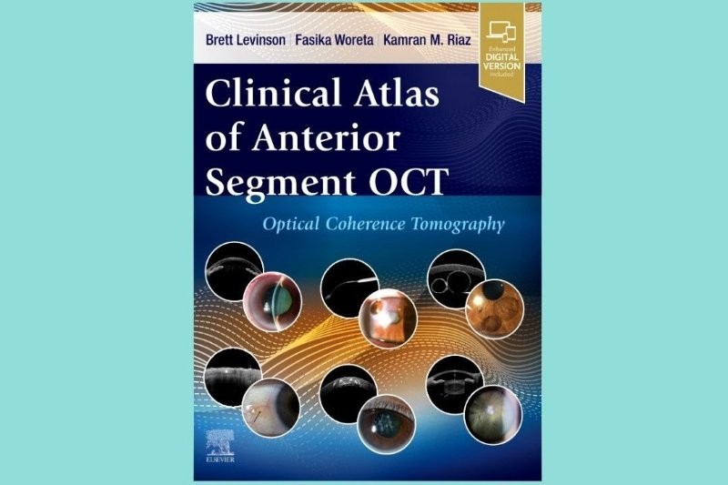
The use of optical coherence tomography (OCT) has become second nature in diagnosing and managing glaucoma and retinal diseases. However, its applications in anterior segment imaging remain underexplored by many eyecare professionals (ECPs). Clinical Atlas of Anterior Segment OCT bridges this knowledge gap by providing a comprehensive guide to the diagnostic and clinical utility of anterior segment OCT (AS-OCT). With its rich repository of high-quality images and detailed commentary, this atlas serves as an invaluable resource for ECPs at all levels.
The book stands out for its exclusive focus on this non-invasive imaging modality, which is crucial for diagnosing and managing corneal, iris, angle and lens abnormalities. Unlike traditional OCT textbooks, which primarily document optic nerve and retinal pathologies, this volume delves deeply into the anterior segment, addressing conditions such as epithelial basement membrane dystrophy and corneal ulcers, angle anatomy, pre- and post-surgical evaluations and scleral contact lens fitting. The book also highlights AS-OCT’s superiority over ultrasound biomicroscopy in visualising some small anterior segment lesions, along with its broader availability in general ophthalmology practices.
The authors spent eight years curating an extensive collection of AS-OCT images, highlighting not only static disease states but also dynamic changes over time. The book is meticulously designed to answer the pivotal question: what can the anterior segment module of OCT do for me?
Organised thematically by anatomy and pathology, the book allows readers to navigate quickly through both common and rare conditions. Each section juxtaposes AS-OCT images with corresponding anterior segment photographs – some with fluorescein photos and topography maps – enabling clinicians to correlate structural findings with ocular pathology. For example, a comparison of epithelial staining and fluorescein pooling in epithelial basement membrane dystrophy exemplifies how AS-OCT enhances diagnostic precision.
The inclusion of clinical pearls in every section underscores the authors’ intent to make this atlas not just informative but also practically useful. The vibrant layout and thoughtful design enhance the reading experience – with bold, colour-coded contents reflecting the practical nature of the book – making it visually appealing and easy to navigate. Special annotations highlight critical features of the anterior segment, ensuring even the most subtle findings are easily identified.
The book also provides a detailed discussion of AS-OCT’s technological advancements. Older models achieved resolutions of 18μm with penetration depths of 3–4mm, while newer iterations reach unprecedented resolutions of 5–7μm. These advancements enable precise quantitative assessments, which are particularly valuable in preoperative planning and postoperative evaluations of corneal surgeries such as penetrating keratoplasty and endothelial keratoplasties as detailed in chapters 13–15. The authors also illustrate how AS-OCT can visualise areas of questionable graft adherence, often revealing details beyond what slit-lamp examination can detect.
Clinical Atlas of Anterior Segment OCT is an essential resource for ECPs and ophthalmic technicians. It not only expands knowledge but also deepens appreciation for the diagnostic power of AS-OCT and fills a critical void in ophthalmic literature. Whether you are exploring AS-OCT’s role in diagnosing corneal pathologies, evaluating anterior chamber angles, or planning refractive surgeries, this book will undoubtedly become a trusted companion.

Vicky Wang is a therapeutically qualified optometrist currently working for Health NZ Te Whatu Ora Auckland.